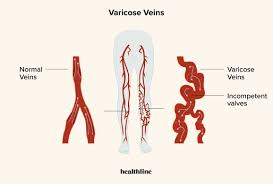
Venous duplex ultrasound, also known as venous Doppler ultrasound, is a non-invasive imaging test used to evaluate the veins in the arms, legs, or other parts of the body. It combines traditional ultrasound imaging with Doppler ultrasound to assess blood flow, detect abnormalities, and diagnose conditions affecting the venous system, such as deep vein thrombosis (DVT), varicose veins, venous insufficiency, or venous reflux.
Procedure
During a venous duplex ultrasound examination, the following steps are typically performed:- Preparation: The patient lies comfortably on an examination table, usually in a supine or slightly elevated position.
- Ultrasound Imaging: A handheld ultrasound transducer, also known as a probe, is placed directly on the skin over the area of interest (e.g., leg) where the veins are being evaluated. The transducer emits high-frequency sound waves that bounce off the veins and surrounding tissues, producing real-time images of the venous system on a monitor.
- Doppler Evaluation: In addition to traditional ultrasound imaging, Doppler ultrasound is used to assess blood flow velocity and direction within the veins. Doppler signals are generated by the movement of red blood cells and are represented graphically on the ultrasound monitor.
- Assessment of Blood Flow: The ultrasound technologist or healthcare provider evaluates the veins for the presence of abnormalities, such as blood clots (thrombi), narrowing (stenosis), or venous reflux (backward flow of blood).
- Compression Testing: In some cases, compression ultrasound may be performed to assess vein compressibility and detect the presence of a deep vein thrombosis (DVT). The veins are gently compressed with the ultrasound probe, and the response of the vein to compression is evaluated.
- Interpretation: The ultrasound images and Doppler waveforms are interpreted by a trained healthcare provider, usually a vascular technologist, vascular surgeon, or radiologist. Abnormal findings are assessed, and a diagnosis is made based on the imaging findings and clinical history.
Indications
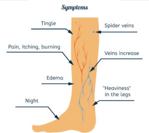
Venous duplex ultrasound may be indicated for patients presenting with symptoms suggestive of venous disease, including:- Swelling or edema in the legs or arms
- Pain or tenderness in the affected area
- Visible varicose veins or spider veins
- Skin changes or discoloration, such as redness or ulceration
- History of blood clots (DVT or pulmonary embolism)
- Chronic venous insufficiency or venous reflux disease
Benefits
AVenous duplex ultrasound offers several benefits for patients suspected of having venous disease, including:- Non-invasive: The procedure is non-invasive and does not involve ionizing radiation or the use of contrast agents, making it safe and well-tolerated by most patients.
- Real-time imaging: Ultrasound provides real-time images of the veins, allowing for immediate visualization and interpretation of any abnormalities.
- High sensitivity: Venous duplex ultrasound has high sensitivity for detecting blood clots (DVT) and other venous abnormalities, allowing for early diagnosis and intervention to reduce the risk of complications such as pulmonary embolism or chronic venous insufficiency.
- Guided interventions: The information obtained from the venous duplex ultrasound can guide further diagnostic testing, such as venography or magnetic resonance venography (MRV), and help determine the need for interventions such as anticoagulation therapy or vein ablation procedures.
Limitations
While venous duplex ultrasound is a valuable diagnostic tool for evaluating venous disease, it has some limitations, including:- Operator dependence: The quality and accuracy of the ultrasound images depend on the skill and experience of the ultrasound technologist or physician performing the procedure.
- Limited visualization: Ultrasound may have limited penetration in obese patients or in areas with deep-seated veins, potentially limiting visualization of the entire venous system.
- Inability to assess smaller vessels: Venous duplex ultrasound may not be able to visualize very small or superficial veins, particularly in patients with extensive varicose veins or venous malformations.


