An echocardiogram, often referred to as an echo, is a noninvasive diagnostic test that uses sound waves to create detailed images of the heart’s structure, function, and blood flow. It is a valuable tool in cardiology and provides essential information for diagnosing and managing various heart conditions.
Procedure
During an echocardiogram, a technician (sonographer) places a small handheld device called a transducer on the chest and moves it around to obtain different views of the heart. The transducer emits high-frequency sound waves that bounce off the heart’s structures and are converted into images on a computer screen. The images show the size, shape, and motion of the heart’s chambers, valves, and surrounding structures.
Types of Echocardiograms
There are several types of echocardiograms, each serving different purposes:
– Transthoracic Echocardiogram (TTE): The most common type of echocardiogram, TTE is performed by placing the transducer on the chest wall to obtain images of the heart from the outside of the body.
– Transesophageal Echocardiogram (TEE): In TEE, a specialized transducer is passed down the throat into the esophagus to obtain closer and more detailed images of the heart’s structures. TEE is often used when higher-resolution images are needed or when TTE is inconclusive.
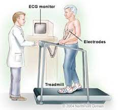
– Stress Echocardiogram: Combines echocardiography with exercise (treadmill or bicycle) or medication (dobutamine) to assess the heart’s function and blood flow under stress conditions.
– Doppler Echocardiogram: Uses Doppler ultrasound to measure the speed and direction of blood flow through the heart’s chambers and valves. It helps evaluate the severity of valve disorders and assess blood flow patterns.
Uses of Echocardiography
Echocardiograms provide valuable information for diagnosing and managing various heart conditions, including:
– Structural Heart Abnormalities: Echocardiography can detect congenital heart defects, valve disorders, cardiomyopathies, and other structural abnormalities of the heart.
– Heart Function: Echocardiograms assess the heart’s pumping function, including measures of ejection fraction, chamber size, and wall motion abnormalities.
– Valvular Heart Disease: Echocardiography evaluates the structure and function of heart valves, including stenosis, regurgitation, and prolapse.
– Cardiac Masses and Tumors: Echocardiograms can detect abnormal growths or masses within the heart, such as tumors or blood clots.
– Pericardial Disease: Echocardiography assesses the pericardium (the sac surrounding the heart) for signs of inflammation, fluid accumulation (pericardial effusion), or constriction.
-Echocardiography is particularly valuable in monitoring heart health after a heart attack or stroke, helping doctors plan appropriate treatments based on how the heart is functioning.
Limitations
While echocardiography is a powerful diagnostic tool, it has some limitations:
– Operator Dependent: The quality of echocardiographic images depends on the skill and experience of the technician performing the test.
– Limited View: Some structures may be difficult to visualize or assess due to body habitus, lung disease, or other factors.
– Invasive Procedures: Transesophageal echocardiography (TEE) requires passing a probe down the throat, which carries some risks and may be uncomfortable for some patients.
Conclusion
Echocardiography is a versatile and valuable diagnostic tool in cardiology, providing essential information about the structure, function, and blood flow of the heart. It is widely used in clinical practice to diagnose heart conditions, guide treatment decisions, and monitor patients’ progress over time.


