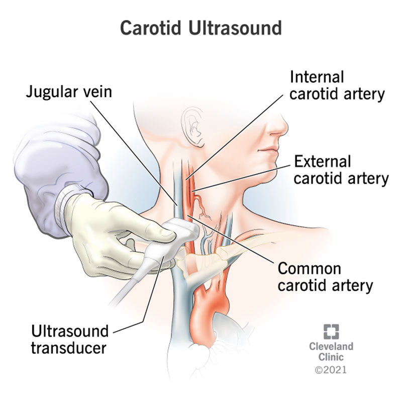
Carotid duplex ultrasound is a non-invasive imaging test used to evaluate the carotid arteries, which are the major blood vessels in the neck that supply oxygen-rich blood to the brain, head, and neck. The carotid duplex ultrasound combines traditional ultrasound imaging with Doppler ultrasound to assess the structure and function of the carotid arteries, including the presence of plaque buildup (atherosclerosis) and any narrowing or blockages that may increase the risk of stroke or transient ischemic attack (TIA).
Procedure
During a carotid duplex ultrasound examination, the following steps are typically performed:
- Preparation: The patient lies comfortably on an examination table, usually in a supine position. A water-soluble gel is applied to the skin over the neck area to facilitate the transmission of sound waves.
- Ultrasound Imaging: A handheld ultrasound transducer, also known as a probe, is placed directly on the skin over the carotid arteries on both sides of the neck. The transducer emits high-frequency sound waves that bounce off the blood vessels and surrounding tissues, producing real-time images of the carotid arteries on a monitor.
- Doppler Evaluation: In addition to traditional ultrasound imaging, Doppler ultrasound is used to assess blood flow velocity and direction within the carotid arteries. Doppler signals are generated by the movement of red blood cells and are represented graphically on the ultrasound monitor.
- Assessment of Blood Flow: The ultrasound technologist or healthcare provider evaluates the carotid arteries for the presence of plaque buildup, narrowing (stenosis), or blockages (occlusions) that may affect blood flow to the brain.
- Measurement of Blood Flow Velocity: Doppler ultrasound measurements are obtained to assess the velocity of blood flow within the carotid arteries and detect any abnormalities, such as turbulence or increased velocities associated with stenosis.
- Interpretation: The ultrasound images and Doppler waveforms are interpreted by a trained healthcare provider, usually a vascular technologist, vascular surgeon, or radiologist. The presence and severity of carotid artery disease are assessed based on the degree of stenosis, plaque characteristics, and blood flow patterns.
Indications
Carotid duplex ultrasound may be indicated for patients presenting with symptoms suggestive of carotid artery disease, including:
- Transient ischemic attack (TIA) or “mini-stroke”
- Stroke or cerebrovascular accident (CVA)
- Symptoms of carotid artery stenosis, such as transient or focal neurological deficits (e.g., weakness, numbness, tingling, or speech disturbances)
- Bruit (abnormal sound) heard over the carotid arteries during physical examination
– History of cardiovascular risk factors, such as hypertension, diabetes, smoking, or hyperlipidemia
Benefits
Carotid duplex ultrasound offers several benefits for patients suspected of having carotid artery disease, including:
- Non-invasive: The procedure is non-invasive and does not involve ionizing radiation or the use of contrast agents, making it safe and well-tolerated by most patients.
- Real-time imaging: Ultrasound provides real-time images of the carotid arteries, allowing for immediate visualization and interpretation of any abnormalities.
- High sensitivity and specificity: Duplex ultrasound has high sensitivity and specificity for detecting carotid artery stenosis and plaque buildup, allowing for accurate diagnosis and risk stratification.
- Early detection and intervention: Carotid duplex ultrasound can detect carotid artery disease at an early stage, allowing for timely intervention and preventive measures to reduce the risk of stroke or TIA.
Limitations
While carotid duplex ultrasound is a valuable diagnostic tool for evaluating carotid artery disease, it has some limitations, including:
- Operator dependence: The quality and accuracy of the ultrasound images depend on the skill and experience of the ultrasound technologist or physician performing the procedure.
- Limited visualization: Ultrasound may have limited penetration in obese patients or in areas with deep-seated arteries, potentially limiting visualization of the entire carotid artery system.
- Inability to assess intracranial vessels: Carotid duplex ultrasound only assesses the extracranial portion of the carotid arteries and does not evaluate intracranial vessels or collateral circulation.
Conclusion
Carotid duplex ultrasound is a non-invasive imaging test used to evaluate the carotid arteries and detect abnormalities such as plaque buildup and narrowing that may increase the risk of stroke or TIA. With its real-time imaging capabilities and high sensitivity, carotid duplex ultrasound plays a crucial role in the diagnosis, risk stratification, and management of carotid artery disease, allowing for timely intervention and preventive measures to reduce the risk of stroke and improve patient outcomes.
A carotid ultrasound, or carotid duplex, is a painless, safe test that uses sound waves to make images of what your carotid arteries look like on the inside. By using the Doppler ultrasound technique, your healthcare provider can also see how well your blood travels through your two carotid arteries , which take blood to your brain.
You may need a carotid artery ultrasound when your healthcare provider wants to look for blood clots or plaque (fat and cholesterol deposits) on your carotid artery walls. These plaque deposits can limit ― and eventually block ― the flow of blood to your brain, face and neck. A blocked carotid artery can cause a stroke.
You may need a carotid ultrasound if you:
- Had surgery on a narrowed artery.
- Need a follow-up on a stent in your carotid artery.
- Are getting periodic checks because your artery was narrow during a previous checkup.
- Have a bruit (unusual sound like a whoosh) your provider could hear through a stethoscope against your artery.
- Have high blood pressure or high cholesterol.
- Had a stroke or transient ischemic attack (TIA).
- Are going to have coronary artery bypass surgery.
- Have diabetes.
- Have stroke or heart disease in your family.
Doppler ultrasound helps your provider look at:
- Blood flow.
- Narrowing or blockage in your artery.
- Malformations you’ve had since birth or tumors.


